Diagnostic technology used the way research trains you to use it: to reduce uncertainty before any irreversible step.
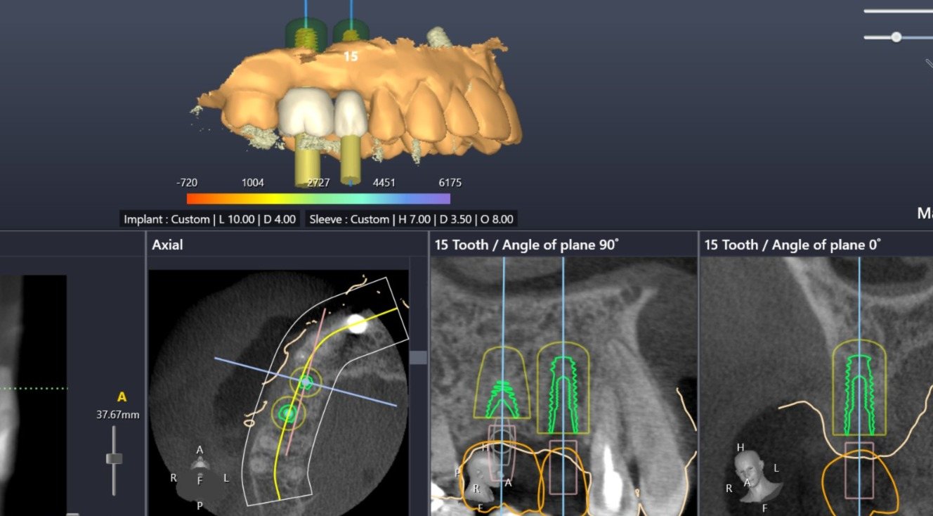
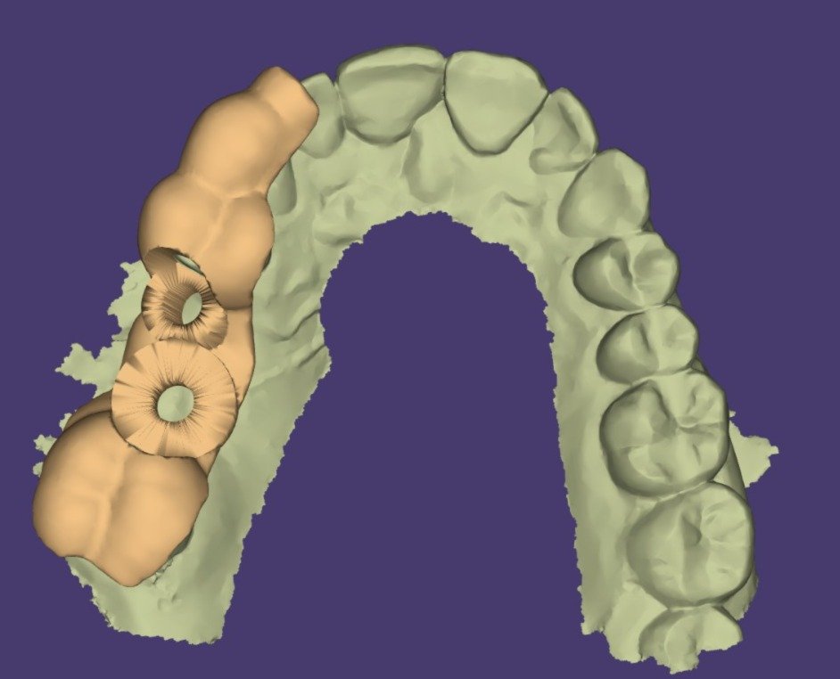
Dr. Meta's academic work depended on precise measurement of bone structure — densitometry, biomechanical testing, tomographic analysis. That same discipline carries into the clinic: digital diagnostics are used to quantify bone volume, plan implant position virtually, and confirm outcomes, rather than relying on estimation.
Cone-beam computed tomography scans, obtained at affiliated imaging centers, are analyzed to build a three-dimensional map of bone volume and density — essential for implant and bone-grafting decisions.
Digital impressions replace conventional molds for many cases, improving comfort and the precision of restorations and aligners.
Lower radiation dose than traditional film, with immediate, enlargeable images for diagnosis and patient education.
Implant position and prosthetic outcome are planned digitally before surgery, reducing surprises during treatment.
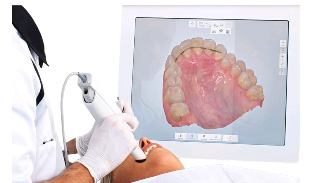
Digital scans and CBCT files can be shared electronically with a patient's home-country dentist or insurer for continuity of care — useful for expatriates coordinating treatment across two countries.